A close look at a small English manuscript

The Fitzwilliam Museum holds an exceptional collection of medieval and Renaissance manuscripts, representing all major schools of European illumination from the ninth to the sixteenth century. In the last decade, hundreds of volumes have benefited from interdisciplinary study undertaken as part of two ongoing projects, Cambridge Illuminations and MINIARE.

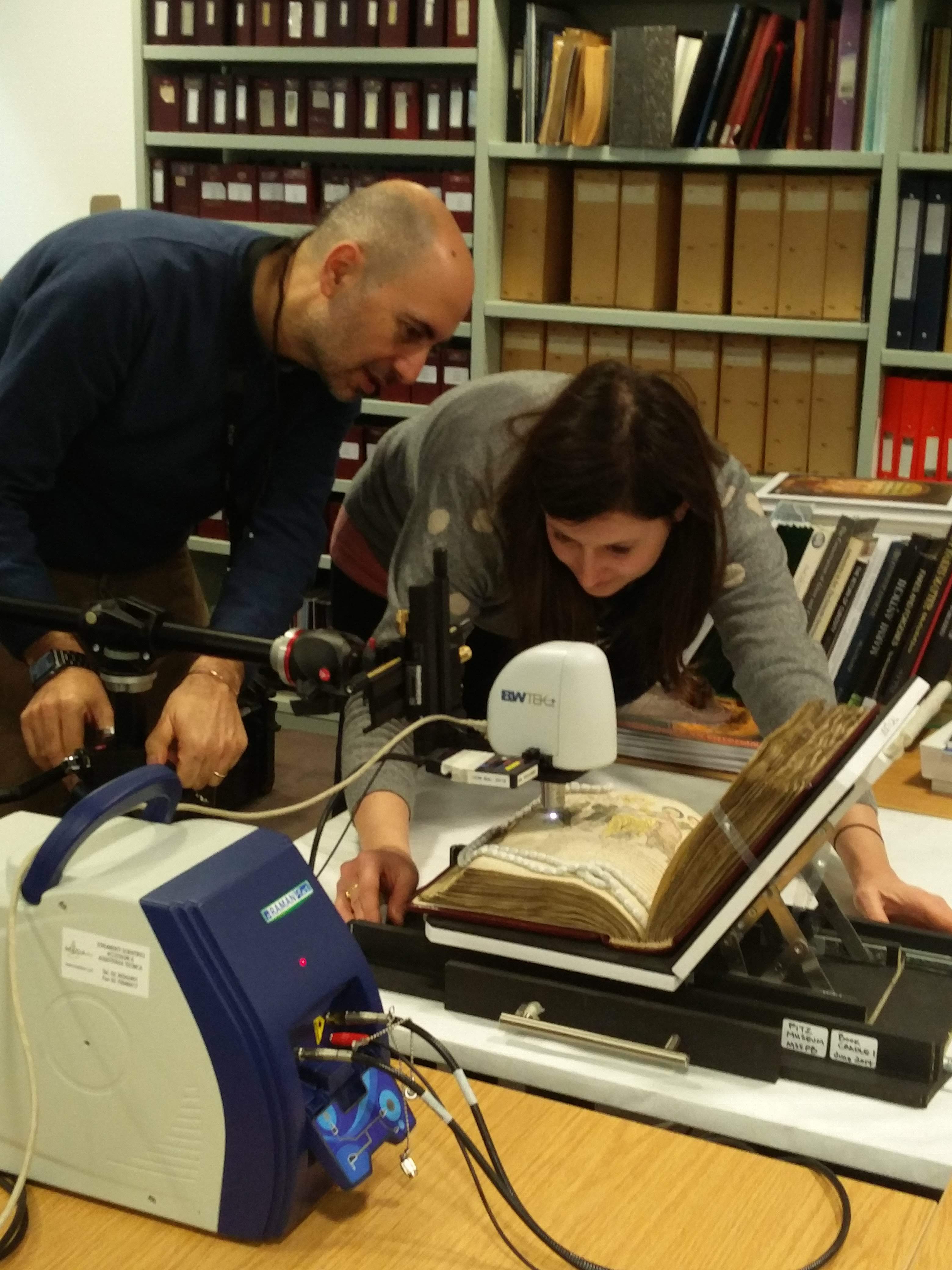
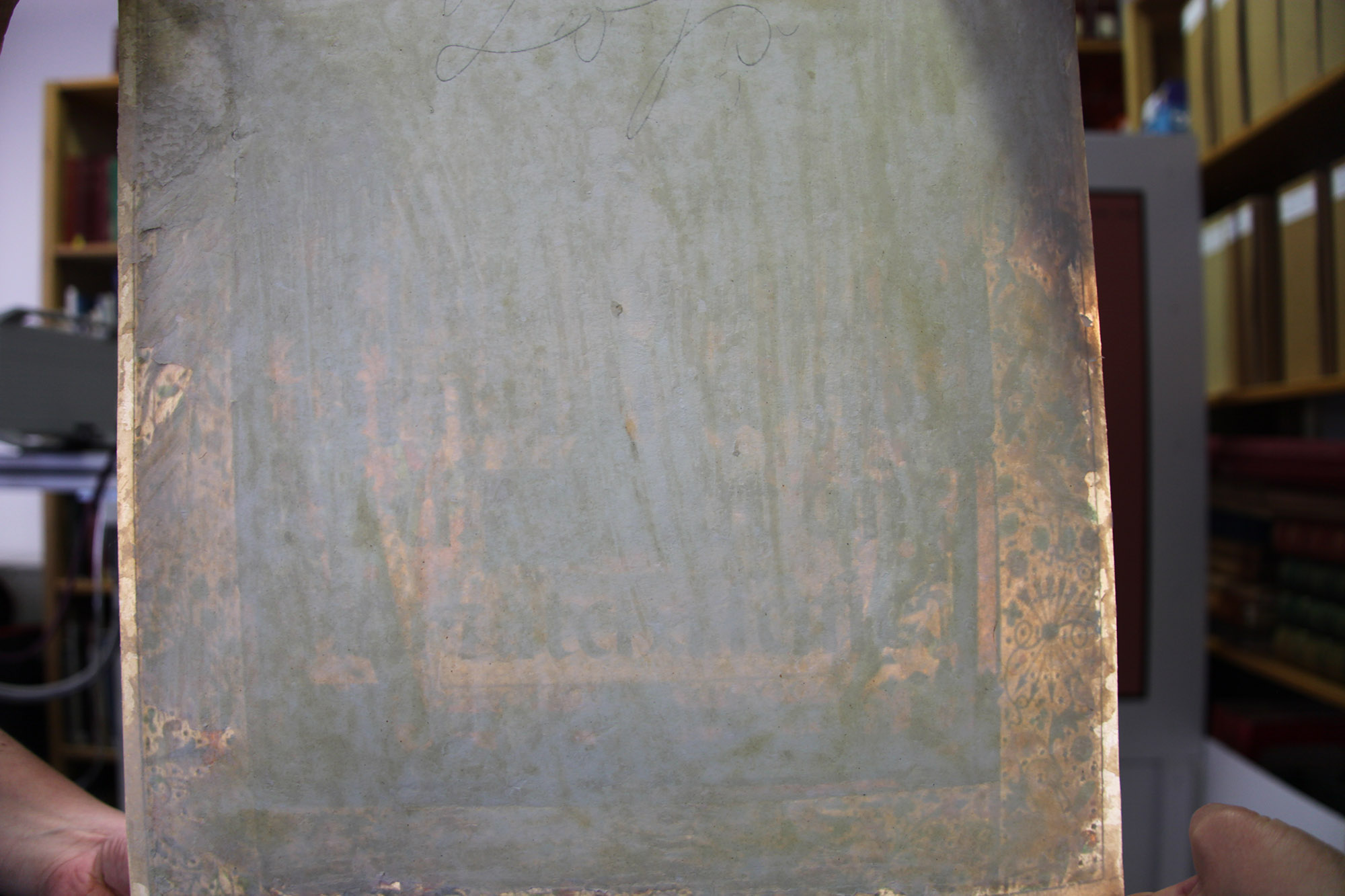
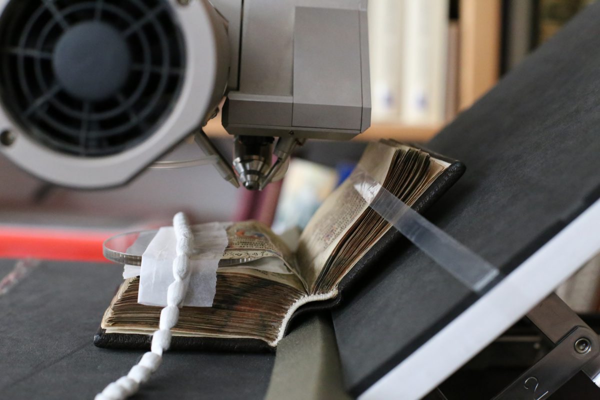
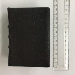
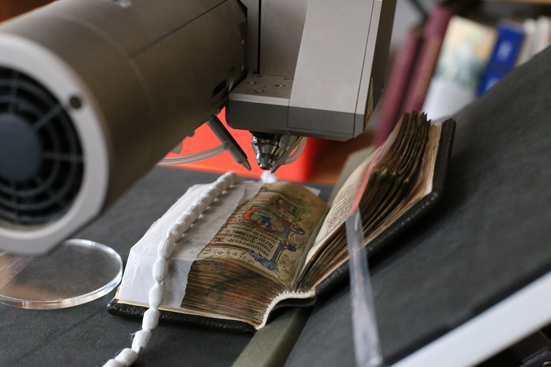
In this context, the Fitzwilliam’s scientific team recently analysed some fifteenth-century English manuscripts in order to investigate the illuminators’ materials and techniques. Among them, we took a close look at a volume which attracted our attention for its dimensions, only about 10 x 7 x 3 cm! (Fig. 1).

Figure 1. MS 2-1967

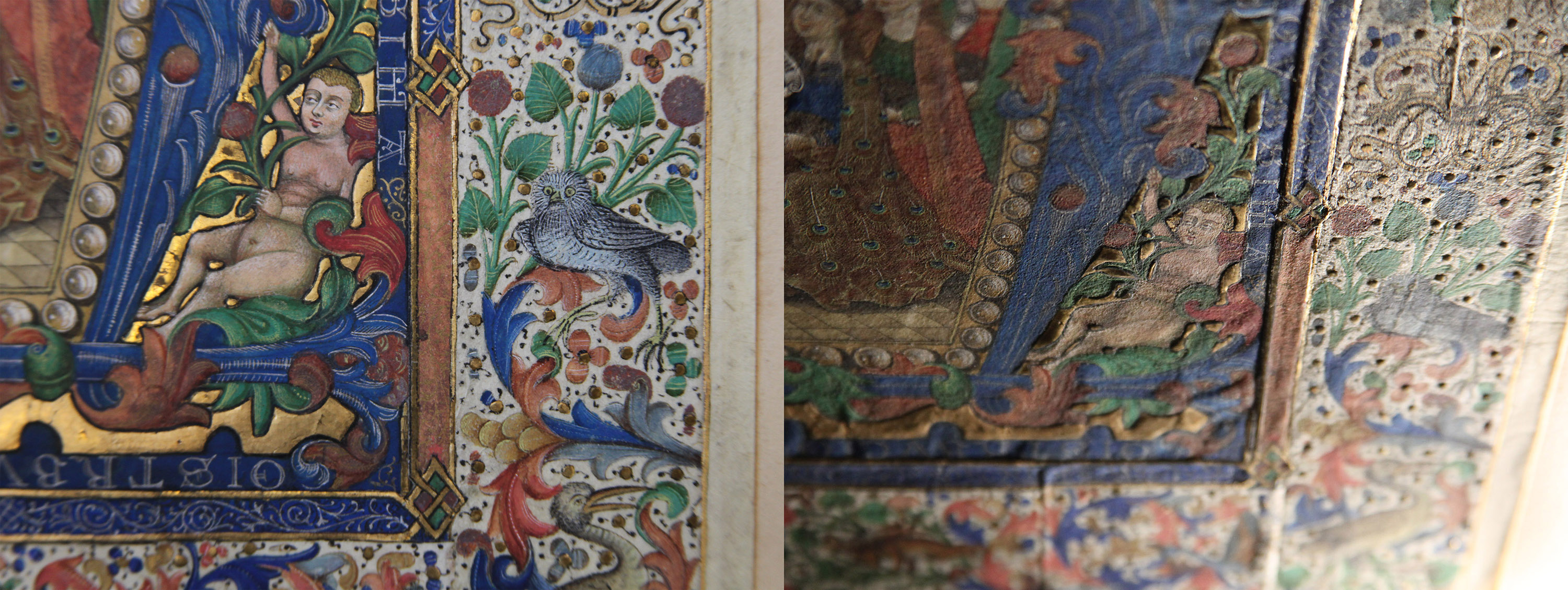
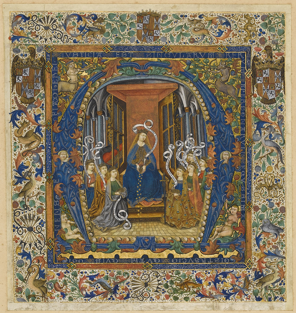
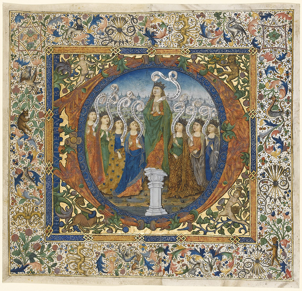
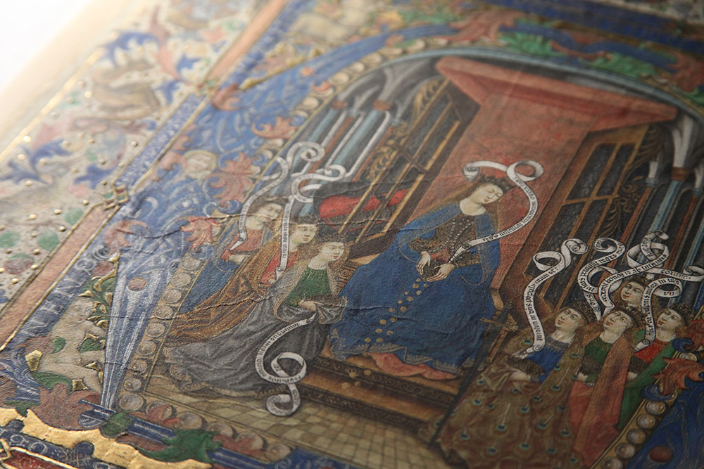
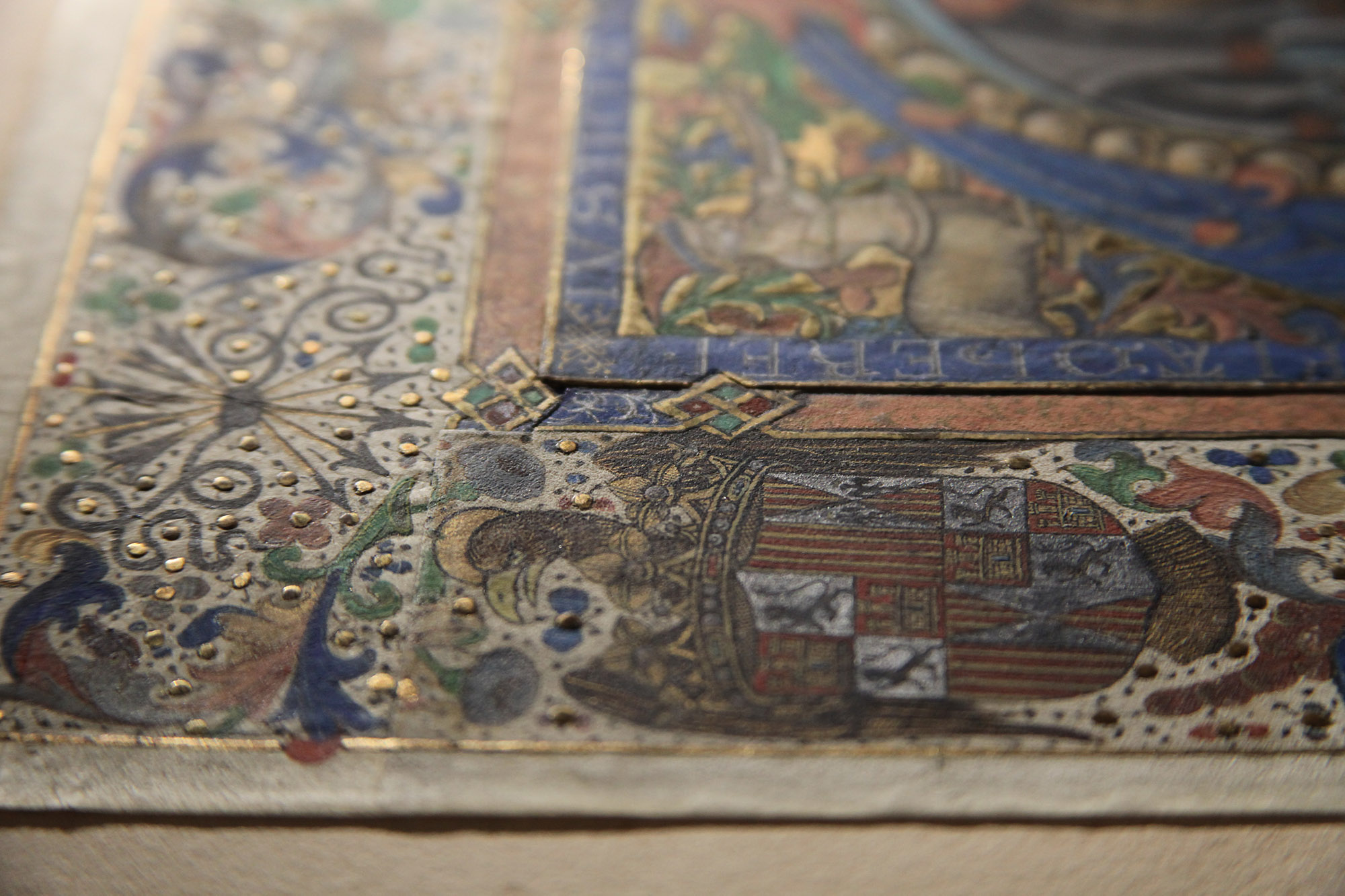
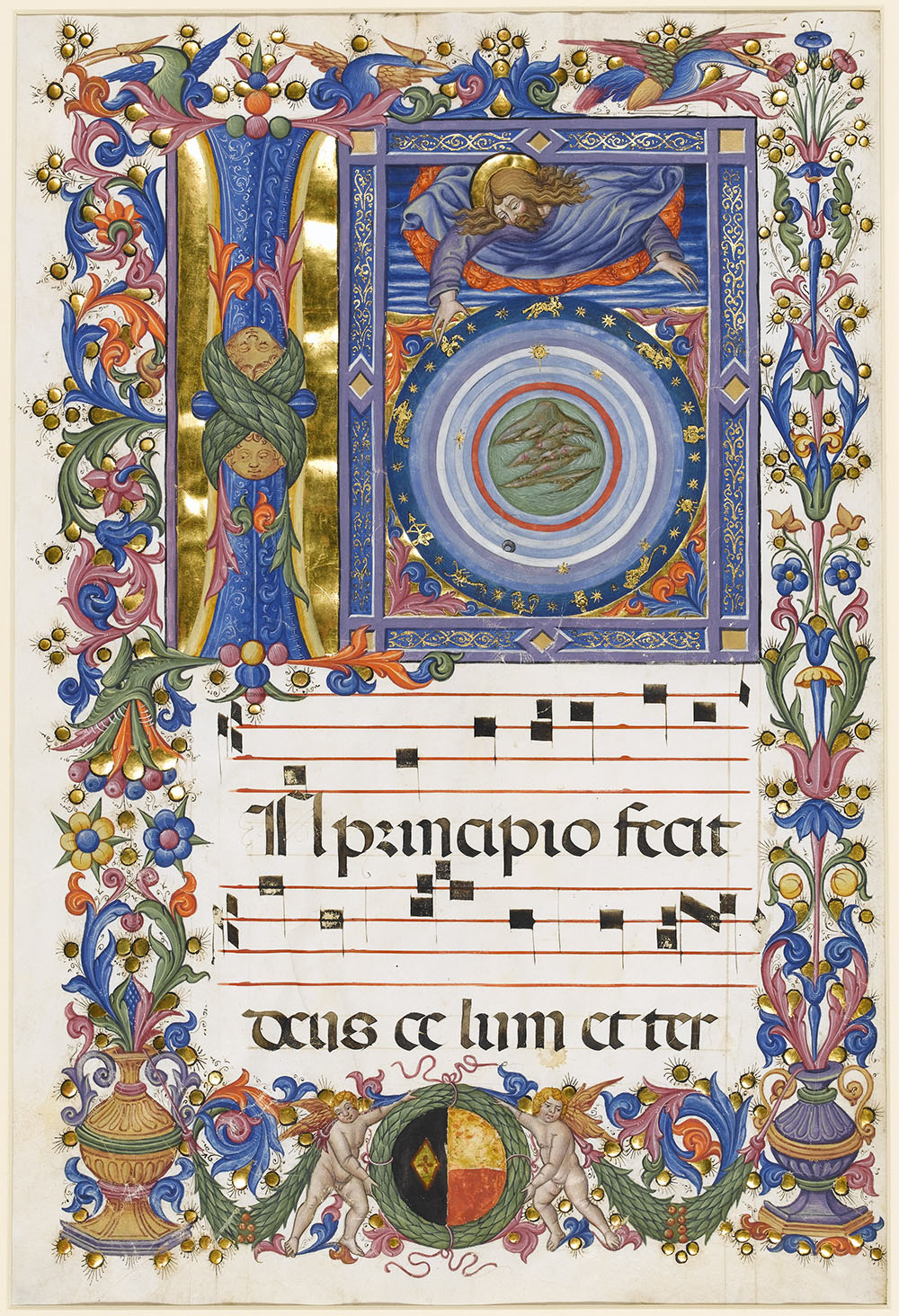
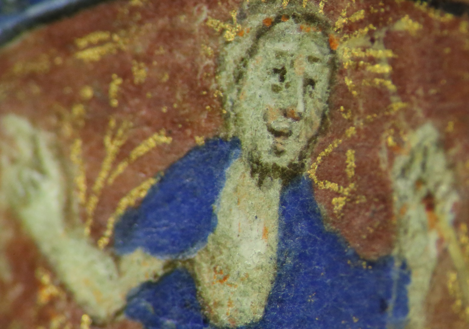
The manuscript (MS 2-1967) is a Book of hours dating to c. 1420, written in Latin on 184 folios of parchment. It contains seven historiated initials, numerous minor decorated initials, pen-work infills of different colours, and borders with golden ivy leaves and coloured acanthus leaves (Fig. 2). Most folios display some level of degradation, in the form of darkening of the red-orange areas and flaking gold leaf, which has significantly changed the original appearance of the decorated borders.

Figure 2. MS 2-1967, fol. 40r (left) and fol. 114r (right). A close look at both folios reveals some design differences in the historiated initials. On fol. 40r, the lower part of the scene extends below the coloured initial and is only partially surrounded by a gilded frame, whereas on fol. 114r, the large initial encloses the miniature and is entirely framed with gold. This may have been a way for a single artist to showcase creativity, or suggest the work of different artists.

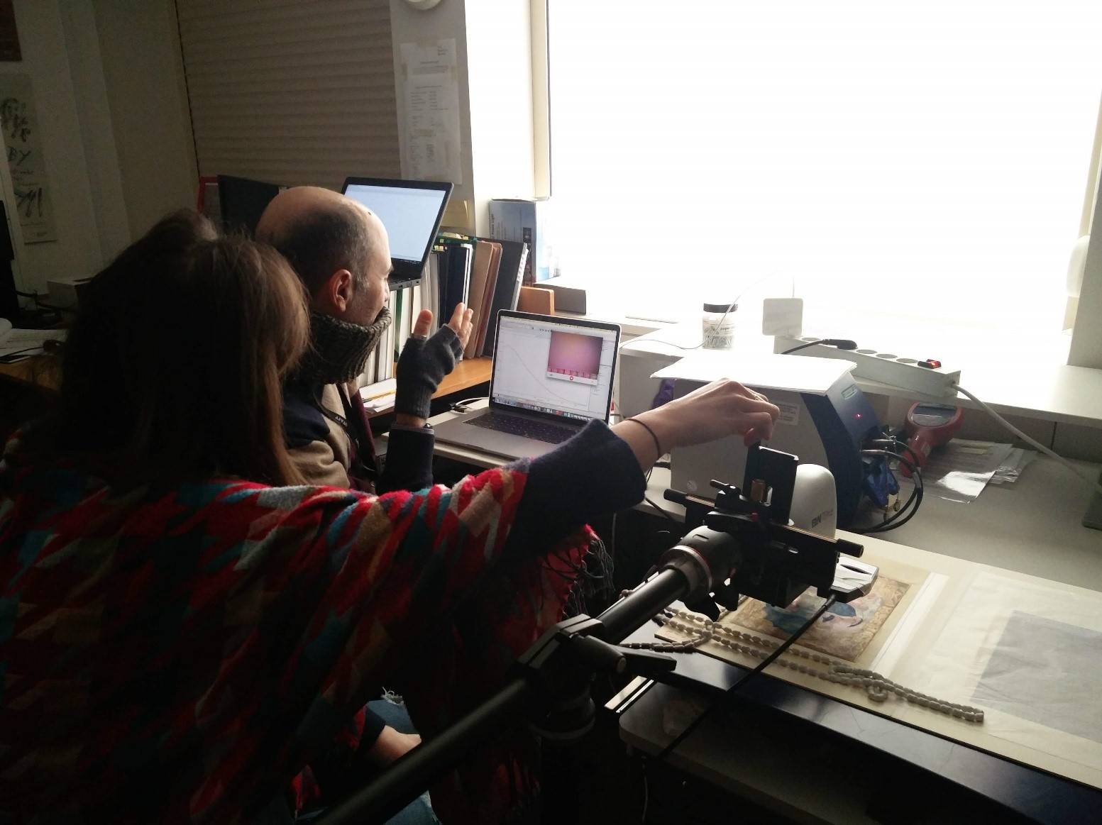
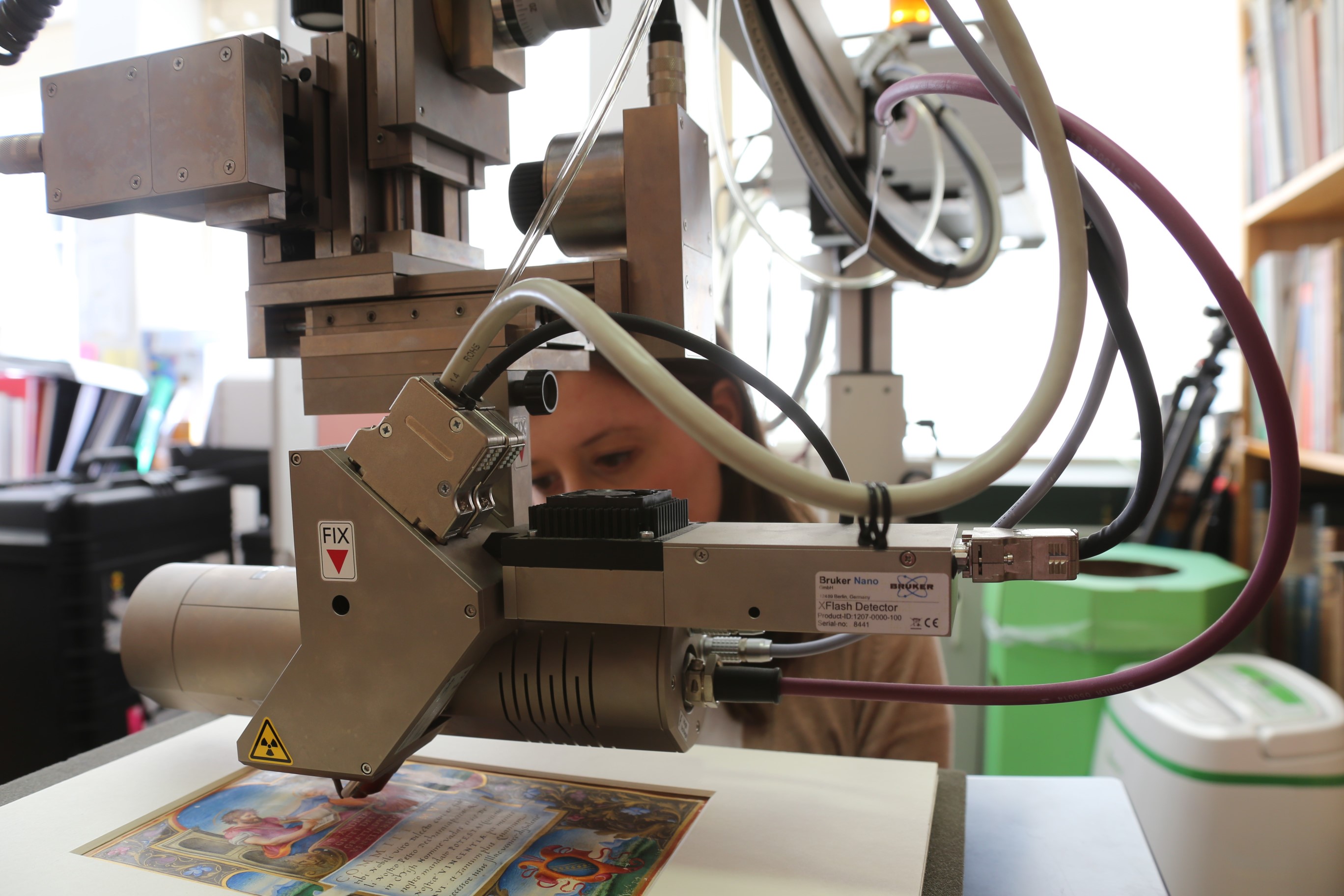
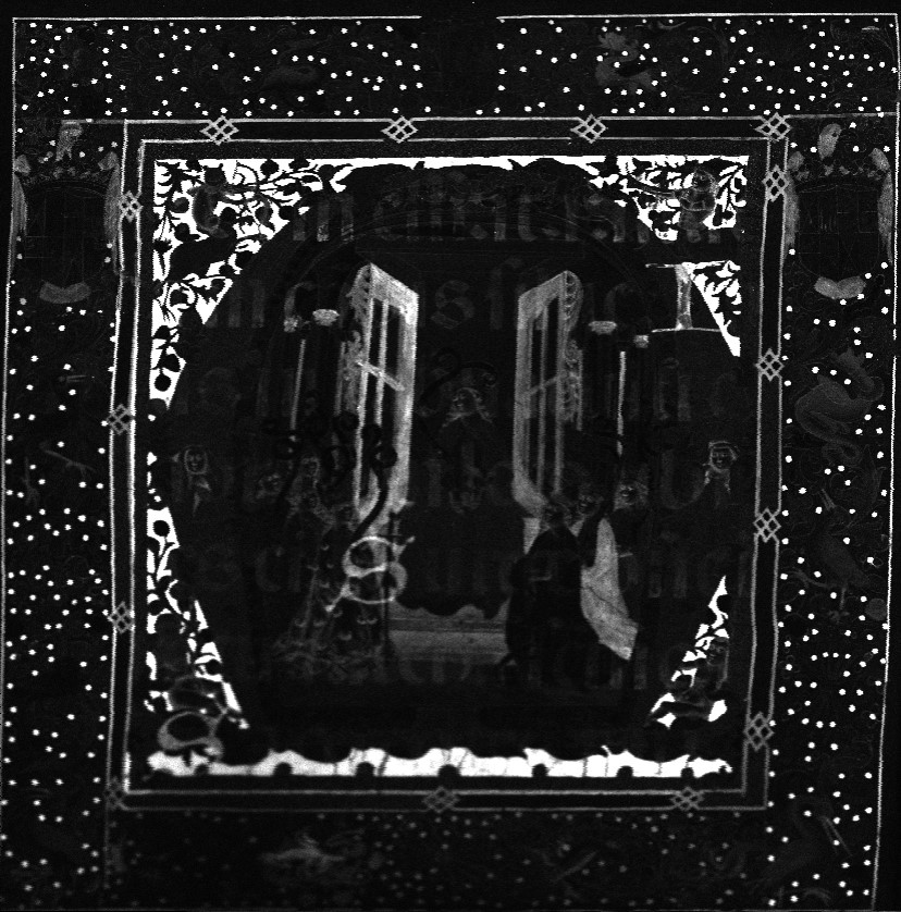
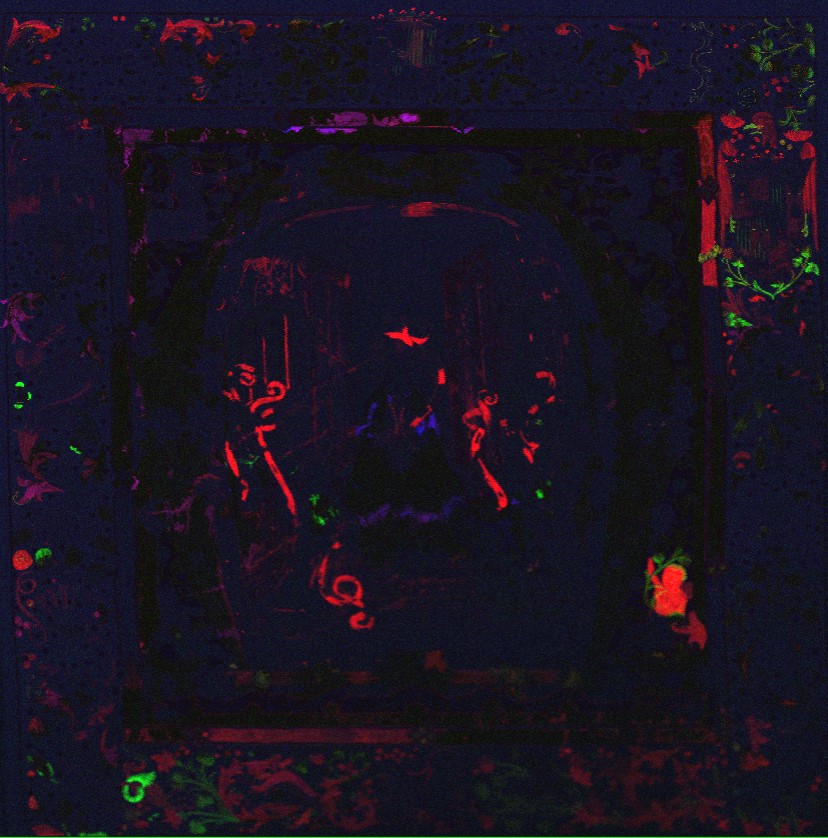
In order to characterise the manuscript’s palette, we examined folios which had been selected by the Keeper of Manuscripts as representative of the  style and colours of the manuscript’s illuminations. We chose a fully non-invasive protocol, i.e. we selected analytical methods that do not require the removal of physical samples or contact with the object’s surface. The analytical protocol included near infrared imaging, reflectance spectroscopy in the ultraviolet, visible, and near-infrared range and X-ray fluorescence spectroscopy (XRF) (Fig. 3).

Figure 3. Preparation for XRF analysis. In order to avoid interference from text and decorations present on the underlying pages, a disc of Plexiglas was carefully laid between the page under analysis and the following one, with a leaded weight securing it in a stable position. Due to the small dimensions and opening characteristics of the manuscript, it was not possible to analyse those areas painted very close to the spine.

The results of the technical investigation revealed a rich palette, which includes lead white, carbon black, vermilion red, and red lead. The latter has often degraded, especially in the borders, and now appears black. An organic red dye was used to paint pink and red passages, whereas a purple dye was employed for lilac pen-work infills surrounding small gilded initials and to rule the pages.

Ultramarine is the main blue pigment used within the illuminations and the text, e.g. to paint all the blue garments and the acanthus leaves. Interestingly, XRF analysis revealed that the ultramarine employed for the small initials within the text contains more calcium than other blue areas analysed. Calcium may derive from calcite, one of the most common minerals associated with the natural stone lapis lazuli, from which ultramarine is made. Its presence may suggest the use of a low-quality ultramarine, prepared or sourced differently than other batches of the same pigment1.

Figure 4. MS 2-1967, fol. 40r. Photomicrograph showing the resurrected Christ. Flesh tones were obtained with a few brown outlines, red dabs, and white highlights; shell gold was used to enrich the red background.

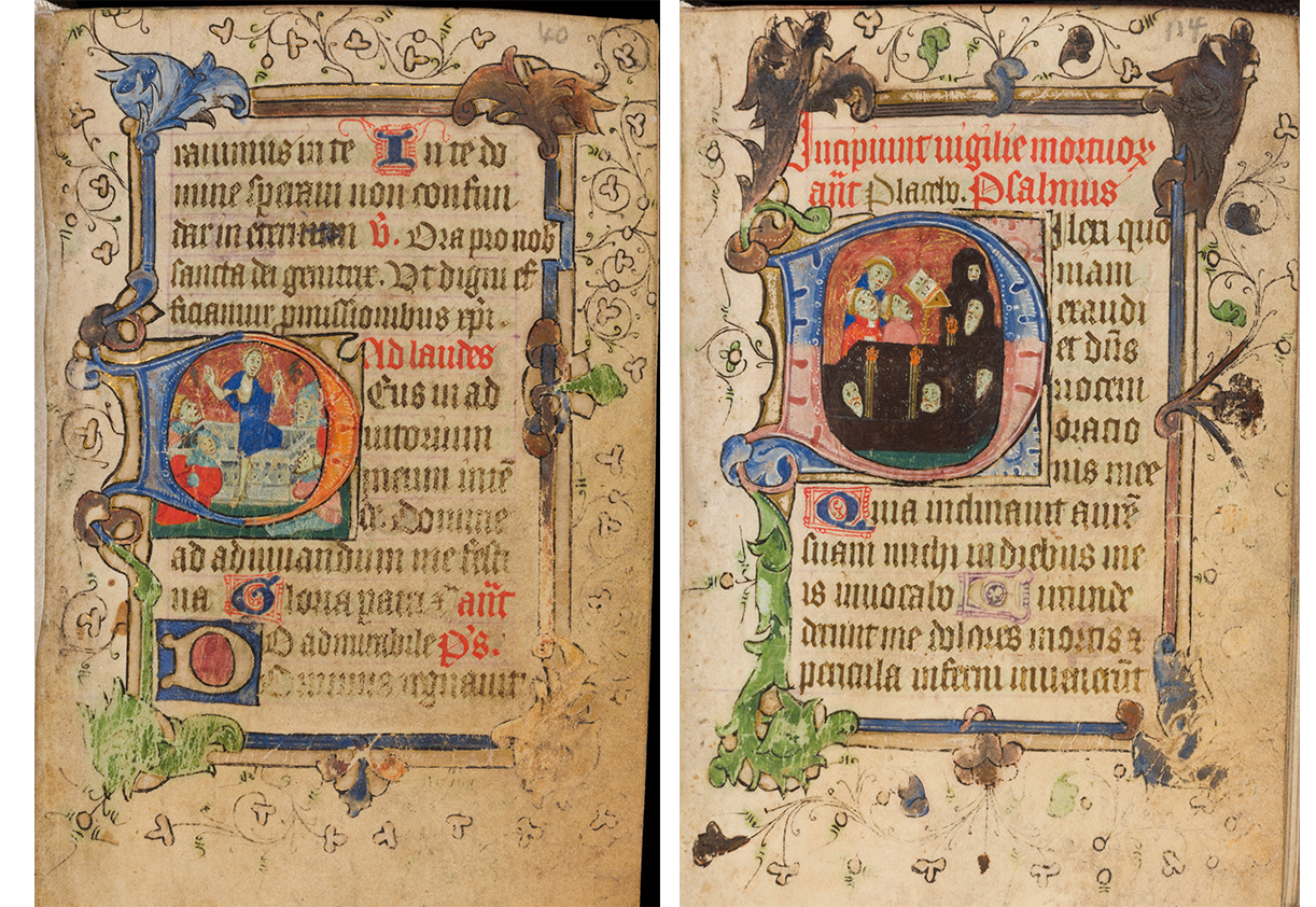
Blue azurite mixed with lead white was found only in small passages, such as the light blue armours of the soldiers witnessing the resurrection of Christ on fol. 40r (see Fig. 2). Azurite was also mixed with an earth pigment to obtain the dark green used in the foreground of all scenes depicted in the historiated initials analysed. An earth pigment, mixed with various compounds, also yielded yellow and brown hues.

Figure 5. MS 2-1967, fol. 40r. Photomicrograph showing one of the soldiers seated outside the Holy Sepulchre looking with astonishment at the resurrected Christ. Brown and yellow hues were obtained using an earth pigment mixed with a copper-based compound and lead white.

A copper-based compound was employed to obtain the bright green leaves of the borders. Its reflectance spectral signature most resembled that of a mineral compound – such as malachite or a copper sulphate –  rather than a synthetic product, such as Verdigris.

Gold was found to be used as shell gold (i.e. powdered gold used as ink or paint) and as gold leaf (i.e. gold beaten into thin sheets), which was laid over a raised white ground. Lastly, iron-gall ink, containing copper and zinc, and red vermillion were used in the text.

Along with the imaging and spectroscopic techniques listed above, microscopic observation helped clarify the illuminator’s painting techniques. Flesh tones were painted using lead white, in addition to a copper-containing compound, an iron-oxide pigment, and small amounts of a calcium-based pigment (such as chalk or gypsum). Outlines and facial features were likely to have been drawn with iron-gall ink; lips, cheeks, and noses were enriched with dabs of red lead, and highlights were then added using lead white.

Figure 6. MS 2-1967, fol. 114r. Photomicrograph showing one of the mourners attending a funeral. Carbon black was used to paint the mourner’s black cloak.

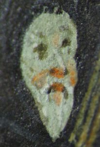
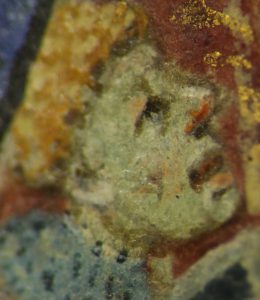
Among the materials detected, two are of particular interest: the copper-containing mineral used for bright green areas, and ultramarine. Both pigments are not commonly encountered in fifteenth-century English manuscripts, which often contain Verdigris and azurite2 instead. Ultramarine remained the standard blue pigment used by illuminators until the late thirteenth century, when it was replaced by azurite, possibly due to the disruption of trade routes between Europe and Asia – the primary source of this pigment – after the disintegration of the Mongol Empire3. The extensive use of precious ultramarine within the manuscript therefore raises questions about the context of its production and the patron’s social status, potentially suggesting a prestigious commission. Additionally, observation under magnification revealed the artist’s ability to portray different expressions and ultimately suggest emotions, such as joy (Fig. 4), astonishment (Fig. 5) or sorrow (Fig. 6), in very tiny faces – they are only a few millimetres long!

Overall, the results of the analyses allowed us to gain insight into the material choices made by a fifteenth-century English illuminator to enrich a book of private devotion. In addition, they will broaden knowledge about English manuscripts produced in that century which have not yet benefited from in-depth examinations.

Even if at first glance the manuscript seemed easy to handle and examine, and the original palette easy to identify, this research taught us that sometimes small objects contain unexpected treasures!

Mila Crippa
Zeno Karl Schindler/MINIARE Fellow
mc2154@cam.ac.uk

References

Osticioli, I. , N.F.C Mendes, A. Nevin, F. Gil, M. Becucci, E. Castellucci, ‘Analysis of natural and artificial ultramarine blue pigments using laser-induced breakdown and pulsed Raman spectroscopy, statistical analysis and light microscopy’, Spectrochimica Acta Part A 73, 2009, 525-531.

Panayotova, S., L. Pereira-Pardo, P. Ricciardi, ‘Illuminator’s Materials and Techniques in Fourteenth-century English Manuscripts’, in Manuscripts in the Making: Art and Science, eds. S. Panayotova and P. Ricciardi, London and Turnhout: Harvey Miller/Brepols, 2017, vol. 1, 46-64.

My first month

If a friend asked me to sum up in brief my first month at work in the Scientific Laboratory of the Fitzwilliam Museum, I would reply, “It has been a crash course in technical analyses of magnificent artworks!” I kick-started the New Year by moving to Cambridge to begin a fellowship as a Research Assistant of the MINIARE Project – Manuscript Illumination: Non-Invasive Analysis, Research and Expertise. Since 2012, the project has carried out scientific investigation of the materials and techniques used to make medieval and Renaissance illuminated manuscripts, while studying the cultural, historical and art-historical context of production.

During the first two weeks, I had the opportunity to catch a glimpse of the rich and diverse collections that the Fitzwilliam Museum houses, crossing the Art History timeline – from ancient Egyptian coffins to French impressionists – simply by walking through the galleries. Among the collections, I became especially acquainted with the Western illuminated manuscripts, which are the focus of in-depth study and conservation for the Department of Manuscripts and Printed Books. Curators and conservators of this Department introduced me into the world of ancient books, making me aware of the conservation needs of unique and fragile artworks, how to choose appropriate temperature, humidity, and lighting conditions, as well as how best to handle the manuscripts before and during the analyses.

Coincidently, the next three weeks fell at the moment visiting researcher Dr Stefano Legnaioli from ICCOM-CNR of Pisa, Italy, came to the Fitzwilliam Museum to carry out analysis of selected objects through Raman spectroscopy. Among the scientific techniques that can be used to examine artworks non-invasively (i.e. without taking samples or touching the artwork’s surface), Raman spectroscopy is one of the most reliable to identify artists’ materials. To enable precise identification, more information was also collected using additional analysis techniques available in the Museum’s Analytical Lab.

Dr Legnaioli and me choosing the best area to carry out Raman analysis on a French manuscript’s folio.

I assisted with the installation of a multi-analytical ‘mobile’ laboratory and helped Dr Legnaioli and the Museum’s Research Scientist, Dr Paola Ricciardi, in performing the analyses. In the brief time available, I was fortunate to be able to work on a ninth-century French manuscript, a lavish copy of the Astronomicum Caesareum printed in the mid-sixteenth century, Corpus Christi College’s Grant of Arms, a group of sixteenth-century Italian Dogali (ducal documents from the Republic of Venice), as well as on some of the Museum’s fan collection.

Dr Legnaioli and me performing Raman analysis on a Venetian Dogale. Monitoring the environmental conditions (high RH and low temperature) is fundamental to ensure the artwork’s conservation. Heavy scarves and gloves are the best equipment to survive!

Our daily protocol included multispectral imaging and spectroscopic analyses: X-ray fluorescence (XRF), reflectance spectroscopy in the ultraviolet-visible-shortwave infrared range (FORS) and Raman. The combination of these analyses revealed underdrawing, changes in the composition, retouching and ultimately the nature of the vibrant and rich colour palettes. The scientific data that we have collected can support other researchers to shed light on the objects’ authorship, style and geographic origin, and ultimately to educate the visitors on how the artefacts were made.

Preparation for XRF analysis of a 16th century Venetian Dogale. In order to ensure the artwork’s safety and to obtain optimal results, the distance between the instrument and the painted surface was carefully evaluated before performing the analysis.

This intensive experience enhanced my knowledge of illuminated manuscripts greatly, and above all, it taught me how to study this type of artefact with a scientific approach. I learned that, even if at first glance a colour palette seems to be easy to identify, analysis can reveal unexpected and unusual results. I understood early on that science cannot answer all the questions that arise at the beginning of research; however, this is a challenge rather than a failure. It may be that we need to find or develop a new protocol, perform other analyses, discuss the problem with curators, or even to leave the question open and unanswered.  I loved doing this cross-disciplinary research because, like a jigsaw puzzle, all the various chemical, physical and historical information can be put together to resolve the manuscripts’ mysteries.

I have just completed the first month of my fellowship at the Fitzwilliam Museum and I look forward to facing new challenges and studying in-depth the art of illumination. With plenty of work to do this year, I am sure there will be new scientific discoveries to write about in future blog posts! I would like to thank the Zeno Karl Schindler Foundation for supporting and funding the MINIARE fellowship, as a remarkable experience for my professional and personal growth.

When fashion transforms the truth

As part of Cambridge’s Festival of Ideas 2017, I took part in a series of behind-the-scenes talks on how we investigate the ‘true’ nature of museum objects. As the event was so successful and attendees showed a lot of interest in the subject, this blog post is aiming to share the story of the objects I discussed on that occasion.

What secrets or lies can we unveil, by which means can we do that, and how far can we go into our understanding of past practices? Some objects lead us into exciting journeys and this is the case of two manuscripts fragments from our collection (MSS 293a and 293b1).

MS 293a (432 x 402 mm)
MS 293b (356 x 376 mm)

These two beautiful and quite peculiar illuminated fragments come from the Royal Monastery of St Thomas in Avila, Spain, and were presented by the Friends of the Fitzwilliam Museum in 1918. By taking different approaches – those of a curator, a conservator and a research scientist – to investigate the truth of these objects, we can retrace their story and reveal some of their secrets.

Historical context and iconography

The description of the image is the first step in our investigation and already gives a lot of information. First we can see that the main part of each image is a large letter: we can identify an ‘M’ and a ‘D’. These are enclosed within composite floral borders containing birds, animals and grotesques, as well as the arms of Castile and Aragon and the devices of the Spanish sovereigns Ferdinand and Isabella, the yoke and the bundle of arrows respectively.

Questions arise already and we start wondering what text these two initials introduced, from which manuscript(s) the images were excised, and what is their context. The catalogue description 2 suggests that the initial ‘M’ would have introduced Psalm 131 (the first psalm for Thursday Vespers) and the initial ‘D’ would have introduced Psalm 114 (the first psalm for Monday Vespers). This type of initial, “shaped and surrounded by densely populated foliage on highly burnished gold grounds, contained within frames inscribed with phrases from the accompanying text”, is, according to our curator Dr Stella Panayotova, “representative of Castilian illumination of the last quarter of the fifteen century which agrees with the internal evidence of the royal arms and devices.” The presence of the arms of Castile and Aragon as well as Ferdinand and Isabella’s devices does indeed give us a lot of information about the time of creation of these images. The two kingdoms of Castile and Aragon were united by this royal couple in 1479; in 1492, the pomegranate (not present here) was added to the royal arms to mark the conquest of Granada. We can therefore conclude that these images were created between 1479 and 1492.

Looking more carefully, we notice the strange layout of the borders. We can see that some of the animals are upside down and some of the drawings are incomplete. We are also aware that this sort of image with such large borders so closely attached to the large initials on all four sides is not commonly seen in illuminated manuscripts. Also, we have two large initials but no other text, so we start asking questions about what happened to these objects and how and why such images came to look the way they do today. At this point we have to start looking ‘beyond’ the image, at the materiality of the object: what is it made of and how can we explain the size and composition of the images?

Conservator’s approach

Last year MS 293a arrived at the conservation studio because it needed some treatment. The fragment was lined with cardboard which was acidic and causing tensions in the parchment. Both of these features were damaging the object.

MS 293a, backing cardboard
MS 293a under raking light

When MS 293a arrived at the conservation studio I followed the usual starting procedure of figuring out what I was dealing with. The catalogue description of the object gave me a good idea of the date of the illumination fragment and its iconography, but very little was written about its materiality.

Using raking light was the most effective observation method to understand the composition of the object. We can see the texture of the surface, giving information about the thickness of the paint layers and the way gold was applied. Raking light also highlights the presence of separate pieces of parchment. So not only is this a fragment from an illuminated manuscript, but it is an assemblage, a collage of several fragments. This explains the very complex completed image, acting as an individual painting.

MS 293a under raking light

Cutting illuminated manuscripts to create new ‘beautiful’ images was a fairly common practice in the 19th century and demonstrates the changing attitudes towards illuminations over time. Mid-19th century revival of interest in Gothic art led to the invasive exploitation of illuminated manuscripts where illuminations were cut and reused in a different context and format: MS 293a was assembled to stand as a painting. It is, as Stella Panayotova writes in the catalogue of the COLOUR exhibition, “a damage inflicted upon illuminated manuscripts, motivated paradoxically, by admiration for them as works of art”.

Backing removal

As mentioned before, the collage was supported by an acidic cardboard. It had been glued with animal glue and stuck firmly onto the board.

The illuminated cutting is painted on parchment which is made of animal skin and is both a very durable but also very reactive material. Parchment becomes stiff over time and reacts hugely to the relative humidity of the surrounding air so tensions can appear when the parchment is forced into a certain shape. My task was to remove the backing and mount the object to keep it safe and stable.

I had to remove the backing board layer by layer in order to reduce the risk of distorting the illumination which could disturb the paint layers. During the process the collage was laid onto a large piece of felt so that the illumination was not crushed. A thin layer of paper was found at the back of the collage and was probably the sheet of paper on which the pieces of the collage were originally adhered. We decided to leave it in place in case removing it would loosen the pieces. However, getting so close to the back of the illumination was intriguing so we looked at the object with transmitted light (now that it was fairly thin) and were able to make out large black Gothic letters. These letters are written on the back of the initial fragment. This was a magical moment and confirmed that the initial belonged to a very large manuscript.

MS 293a backing removal
MS 293a through transmitted light after backing removal

We had already so much information and could be fairly confident in attributing the illuminated initials to a Choir Psalter, written in Latin and originating from Avila in Spain in the late 15th century.

We also found out via observations under natural light and raking light that the collages were made of 8 fragments (MS293a) and 3 fragments (MS293b) respectively. Our curator was fairly confident in attributing the floral borders to the same illuminated manuscript as the initials. We could now go further in our understanding of the object and being able to reveal how the ‘artist’ who made the collage achieved the final composition. This is where our research scientist comes in.

Scientific approach

Collaboration with Dr Paolo Romano and Claudia Caliri from the LANDIS Laboratory in Catania (LNS-INFN and IBAM-CNR), allowed us to analyse MS 293a with a cutting-edge macro-XRF scanner, which they shipped here all the way from Italy! The spectral images obtained were interpreted by our research scientist and here is what they revealed:

The elemental map for iron (Fe-K) shows the presence of iron in various areas. The obvious place where we can see iron is in the gilded areas because iron is a constituent of Armenian bole, which would have been used as the ground layer for the application of gold leaf. However, we can also see what appear to be letters throughout the centre of the image. By flipping the image over (see image below) and adjusting its contrast we realise that these are indeed letters, written in iron-gall ink on the other side of this parchment page. Careful observation allows us to start reading the text hidden behind the collage.

MS 293b, elemental map for iron (Fe-K) flipped left-to-right

The false-colour elemental map for zinc, chromium and cobalt (in red, green and blue, respectively ) reveals the presence of elements, all of which are characteristic of ‘modern’ pigments. Combining this information with other analytical data, we can identify the presence of zinc white, cobalt blue, barium chromate and a chromium-based green, all of which were first manufactured in the first half of the 19th century. This information, together with the absence of any pigments manufactured after 1850, suggests that the collage was assembled, and the image retouched, probably in the 1840s, in line with the 19th-century practice of excising illuminations from manuscripts to create new art objects.

MS 293a, false-colour elemental map for zinc, chromium and cobalt
Conclusion

Collaboration between curator, scientist and conservator helped to retrace the origins and the story of these collages and to document them. These objects are new valuable objects, made of ancient valuable fragments. We won’t undo the collages because they document historic practice on top of being a new artwork. Besides, there is currently no parent manuscript so re-uniting the fragments is not possible – and if it was, digital methods could allow us to reunite fragments from all over the world by use of online digital tools, for example the IIIF (International Image Interoperability Framework). Separating the pieces could also damage the parchment and the paint layers, and having individual fragments without a specific place or use for them wouldn’t be appropriate.

Revealing the truths of an object is so important because we can’t undo what has been done for various reasons explained previously. However, we can document the object and share the information. One day, perhaps, through sharing information, we will find more related fragments that will feed into our understanding of the object itself and the historical practices involved and we may even be able to start reconstructing the illuminated manuscript that was once proudly standing in the Royal Monastery of St Thomas in Avila.


MS 197 (578 x 400 mm)

This cutting from our collection gives an idea of the scale of a Choir Plaster and how the fragments of the collage could have been laid out on a page. This is MS 197, from a 15th-century Italian Choir Book (see catalogued information’s here). We can see the large initial, the floral borders, the coat of arm and large black manuscript letters.


With special acknowledgement to Dr Paola Ricciardi, Fitzwilliam Museum’s Research Scientist, for her input with scientific research and imaging and for her help with co-writing this blog post.