Cataloguing and Digitising the Jim Murrell Archive

Jim Murrell

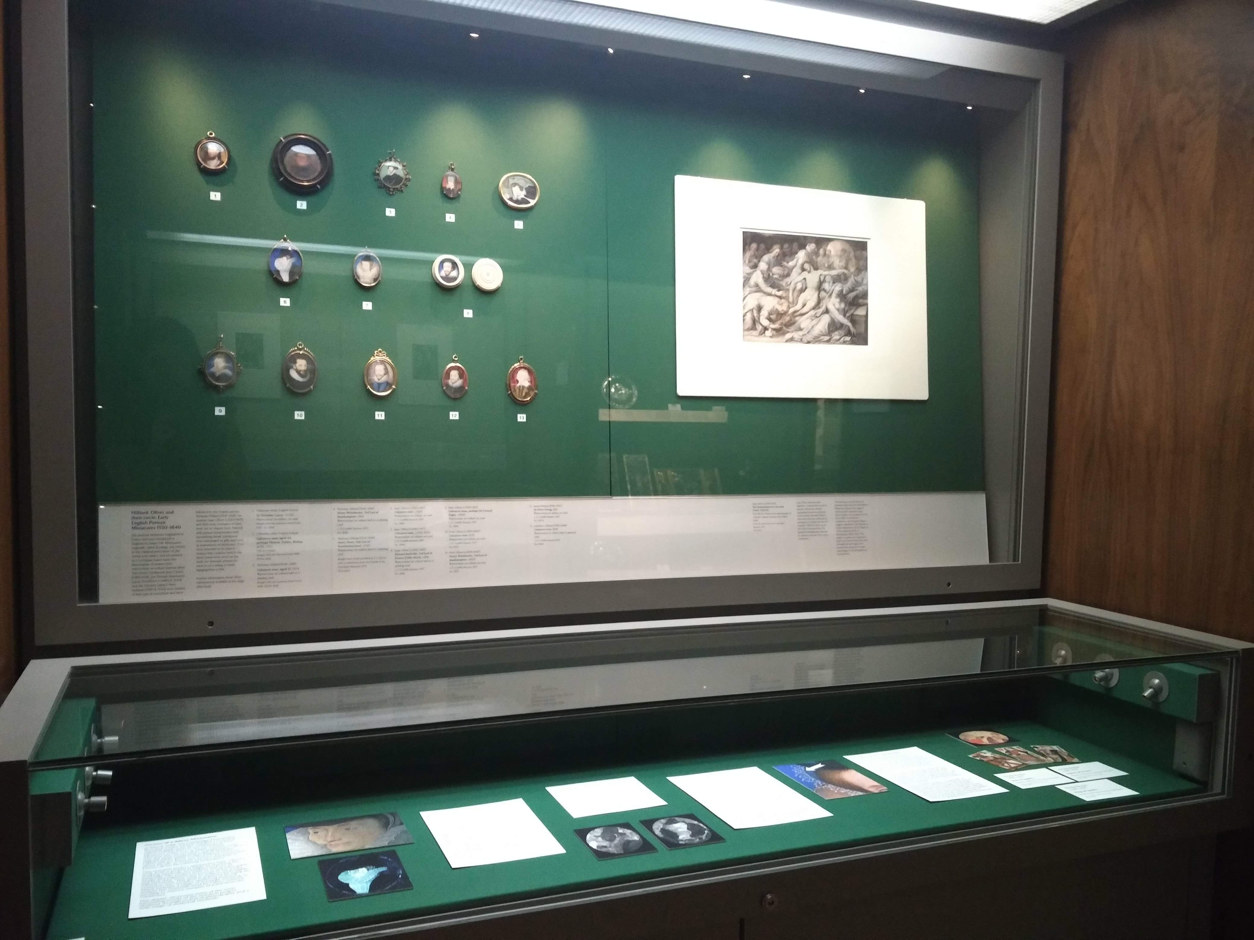
For anyone working on the topic of early modern portrait miniatures, 2019 was an exciting year, seeing the fruition of much new research in exhibitions and publications, including the large exhibition Elizabethan Treasures: Miniatures by Hilliard and Oliver (21 Feb 2019 – 19 May 2019), which introduced a new generation to this art form at the National Portrait Gallery, London; Elizabeth Goldring’s much-awaited biography, Nicholas Hilliard: Life of an Artist, (New Haven, London: Yale University Press, 2019); and Cambridge’s very own focused display, Secrets of a Silent Miniaturist: Technical Analysis of Isaac Oliver’s Miniatures at the Fitzwilliam Museum. In Cambridge, work on miniatures continues with the technical analysis of Oliver’s work and, as part of this project, the digitisation of the hitherto largely unexplored archive of Jim Murrell (1934–1994), housed at the Hamilton Kerr Institute (HKI).

A selection of the Fitzwilliam Museum’s miniatures, show-casing some of the recent discoveries on the work of Isaac Oliver, in the Rothschild Gallery of Medieval & Renaissance Art (Gallery 32)

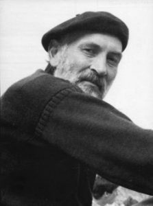
Vernon James Murrell, known as Jim, was a conservator of miniatures and wax objects at the Victoria and Albert Museum, London (V&A) from 1961 until his retirement in 1994. Murrell worked on the V&A’s National Collection of portrait miniatures as well as examples in private and other public international collections. He wrote and contributed towards a number of key publications, in which he shared his technical knowledge of miniatures.  These include John Murdoch et al., The English Miniature (New Haven, London: Yale University Press, 1981); Roy Strong and V. J. Murrell, Artists of the Tudor Court: The Portrait Miniature Rediscovered, 1520–1620 (London: V&A, 1983); and The Way Howe to Lymne: Tudor Miniatures Observed (London: V&A, 1983). His edition of Edward Norgate’s seventeenth-century treatise Miniatura, or The Art of Limning, co-authored with Jeffrey M. Muller, was published posthumously (New Haven, London: Yale University Press, 1997). Murrell’s work pioneered the technical study of miniatures and the communication of his findings to non-specialist audiences, and continues to be used today by art historians and new audiences.

A selection of the two-hundred-plus files, books and boxes in the Jim Murrell Archive at the Hamilton Kerr Institute

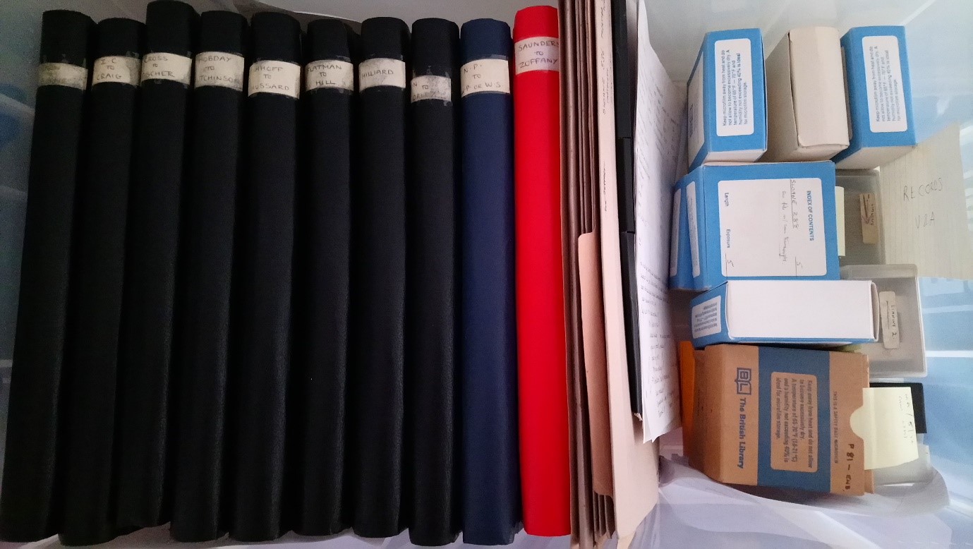
The archive contains Murrell’s notes, sketches, slides, and lectures, most of which have not yet been published, as well as secondary reading materials.It  reveals adesire to understand how miniatures were made, the materials and techniques which were used to create them, and Murrell’s curiosity concerning the technical interest in miniatures in early modern Britain. His notes reveal how artists created miniatures, what pigments were employed for the paints, and how the artist applied the paint to the support. Murrell transcribed copies of historical manuscripts and annotated them to indicate where recipes were unique, had been copied from other treatises, and where they offered a variation on existing knowledge. These annotations highlight the ways in which information circulated amongst artists, patrons and other interested readers in early modern England. Information about the painting of portrait miniatures can also be found within a variety of written materials on other topics, including commonplace books, books of coats of arms and heraldry, and books on plants.

The archive was donated to the HKI by Jim’s wife, Ann Murrell Ballantyne, a restorer of medieval wall paintings, in June 1999. Access to the archive is currently greatly limited but plans are afoot to create an archive centre at the HKI. In the meantime, however, and for those who are not able to travel to the HKI, it is hoped that the digitised version will soon be available online, providing access to reproductions of Murrell’s notes and sketches.1

Lindsey Cox digitising the Jim Murrell Archive at the Hamilton Kerr Institute

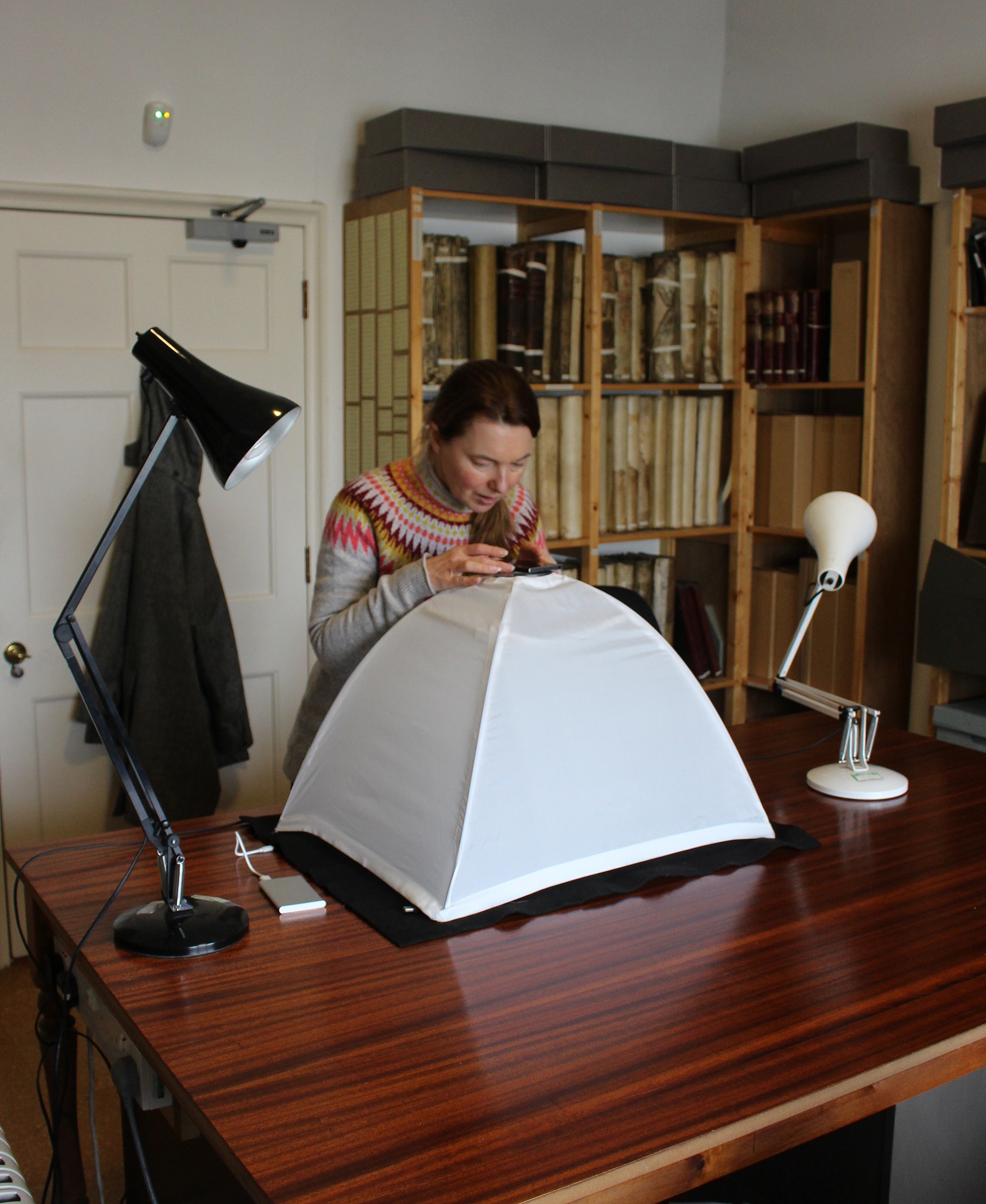
Between April and December 2019, I digitised over five thousand images from the archive. The images were captured using DocScan, a free mobile phone scanning app which was installed on a Sony Xperia XA1. Docscan works with the mobile camera and does not require a flatbed scanner.  This would make it a good option for researchers visiting archives where no scanner is available or where scanning may damage the original. Often, the DocScan app could detect the outlines of a page and suggested where to crop and edit the images before saving to the phone. I found that this function worked better with pages of typed text than with notebooks of faint, pencilled sketches or notes. Once the images had been captured and edited, they were saved as PDF files and transferred to a computer. This made it easier to view the image to check for focus and cropping. Sometimes images needed to be taken again to ensure legibility, which was the main priority of this project. Digitising the archive will not only increase accessibility, but also help decipher Murrell’s notes: his script is tidy but sometimes very small, and is therefore easier to read once it has been magnified on the computer. Digitisation will also help to ensure the longevity of Murrell’s knowledge, should anything ever happen to damage the original material.

Image showing a page from one of Murrell’s notebooks

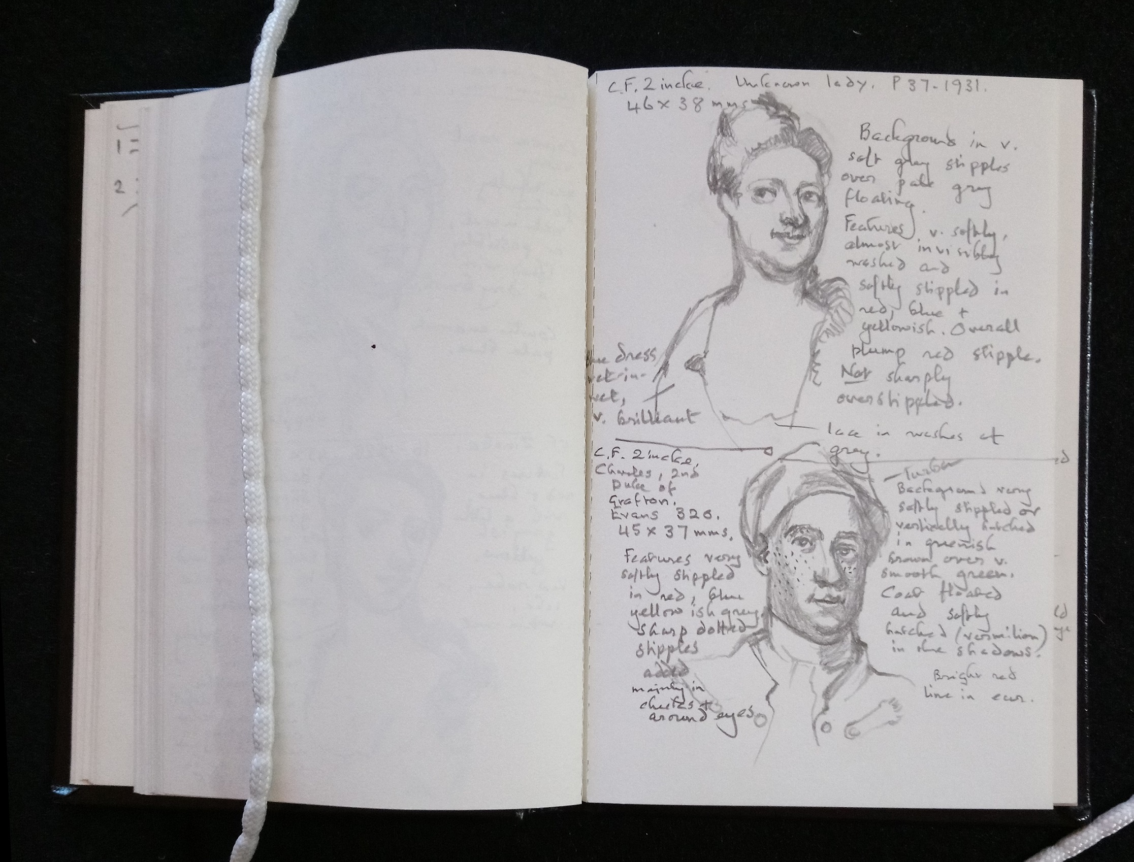
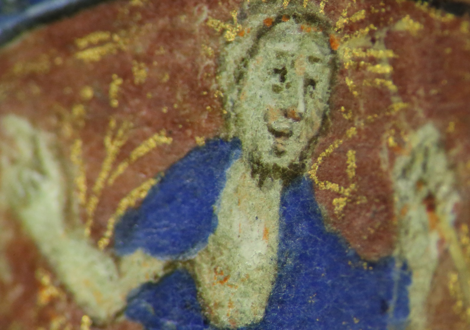
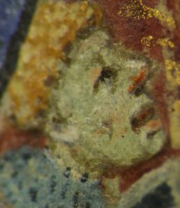
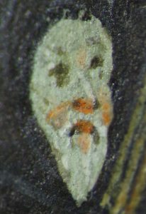
The image above shows a digitised page from one of Murrell’s notebooks in which he has included notes and sketches from the V&A collection of portrait miniatures. The upper image shows Murrell’s sketch of an unknown lady painted by the enamel miniature artist, Christian Friedrich Zincke, c. 1705–1745, 46 mm x 38 mm (P.37-1931). With the benefit of viewing the original painting under magnification combined with his technical knowledge of how these works were created, Murrell has noted that the sitter’s blue dress was painted ‘wet-in-wet’, a painting technique in which paint layers are applied one after the other, before the previous layerhas dried. This technique is used to create a very smooth appearance with no visible brushstrokes. Murrell also noted this highly finished effect in the pale grey ‘floating’ background of the miniature, and the almost invisible washes laid down to create the features of the figure. Below this in Murrell’s notebook is a sketch of a second miniature by Zincke: Charles, 2nd Duke of Grafton, c. 1730, enamel on metal, 45 mm x 37 mm (Evans 320). Again, Murrell’s close observation of the work reveals the techniques whereby Zincke achieved his smooth effects. The background is noted as ‘very softly stippled’,a technique of using small dots or short strokes of the paintbrushwhereas  vertical hatching (closely drawn lines) and ‘sharp dotted stipples’ are used by the painter to model the sitter’s face. The different sorts of marks are clearly represented in Murrell’s sketch. The notebook contains further sketches and notes on miniatures by Zincke and his contemporaries.

The archive now exists in its original state and, largely, as a series of digitised files. With further funding, it will be possible to make these digitised files available for public viewing online. It is hoped that providing the Murrell archive with an online presence will provide an ongoing legacy and foster  the revival of interest in miniatures. The funding for the digitisation work undertaken so far was granted by the British Academy Small Research Grant scheme and the Fitzwilliam Museum’s Marlay Group, as part of an ongoing technical research project on Isaac Oliver.

If you want to know more or contribute to the project, please get in touch by emailing portraitminiatures@fitzmuseum.cam.ac.uk.

Bibliography

DocScan http://docscan.ifunplay.com/

Elizabeth Goldring, Nicholas Hilliard: Life of an Artist, (New Haven, London: Yale University Press, 2019)

Jeffrey M. Muller and Jim Murrell, Edward Norgate: Miniatura, or, the Art of Limning (New Haven, London: Yale University Press, 1997)

Jim Murrell, The Way Howe to Lymne: Tudor Miniatures Observed (London: The Victoria and Albert Museum, 1983)

Roy Strong and V. J. Murrell, Artists of the Tudor Court: The Portrait Miniature Rediscovered, 1520–1620 (London: V&A, 1983)

John Murdoch, Jim Murrell, Patrick J. Noon and Roy Strong, The English Miniature (New Haven, London: Yale University Press, 1981)


A close look at a small English manuscript

The Fitzwilliam Museum holds an exceptional collection of medieval and Renaissance manuscripts, representing all major schools of European illumination from the ninth to the sixteenth century. In the last decade, hundreds of volumes have benefited from interdisciplinary study undertaken as part of two ongoing projects, Cambridge Illuminations and MINIARE.

In this context, the Fitzwilliam’s scientific team recently analysed some fifteenth-century English manuscripts in order to investigate the illuminators’ materials and techniques. Among them, we took a close look at a volume which attracted our attention for its dimensions, only about 10 x 7 x 3 cm! (Fig. 1).

Figure 1. MS 2-1967

The manuscript (MS 2-1967) is a Book of hours dating to c. 1420, written in Latin on 184 folios of parchment. It contains seven historiated initials, numerous minor decorated initials, pen-work infills of different colours, and borders with golden ivy leaves and coloured acanthus leaves (Fig. 2). Most folios display some level of degradation, in the form of darkening of the red-orange areas and flaking gold leaf, which has significantly changed the original appearance of the decorated borders.

Figure 2. MS 2-1967, fol. 40r (left) and fol. 114r (right). A close look at both folios reveals some design differences in the historiated initials. On fol. 40r, the lower part of the scene extends below the coloured initial and is only partially surrounded by a gilded frame, whereas on fol. 114r, the large initial encloses the miniature and is entirely framed with gold. This may have been a way for a single artist to showcase creativity, or suggest the work of different artists.

In order to characterise the manuscript’s palette, we examined folios which had been selected by the Keeper of Manuscripts as representative of the  style and colours of the manuscript’s illuminations. We chose a fully non-invasive protocol, i.e. we selected analytical methods that do not require the removal of physical samples or contact with the object’s surface. The analytical protocol included near infrared imaging, reflectance spectroscopy in the ultraviolet, visible, and near-infrared range and X-ray fluorescence spectroscopy (XRF) (Fig. 3).

Figure 3. Preparation for XRF analysis. In order to avoid interference from text and decorations present on the underlying pages, a disc of Plexiglas was carefully laid between the page under analysis and the following one, with a leaded weight securing it in a stable position. Due to the small dimensions and opening characteristics of the manuscript, it was not possible to analyse those areas painted very close to the spine.

The results of the technical investigation revealed a rich palette, which includes lead white, carbon black, vermilion red, and red lead. The latter has often degraded, especially in the borders, and now appears black. An organic red dye was used to paint pink and red passages, whereas a purple dye was employed for lilac pen-work infills surrounding small gilded initials and to rule the pages.

Ultramarine is the main blue pigment used within the illuminations and the text, e.g. to paint all the blue garments and the acanthus leaves. Interestingly, XRF analysis revealed that the ultramarine employed for the small initials within the text contains more calcium than other blue areas analysed. Calcium may derive from calcite, one of the most common minerals associated with the natural stone lapis lazuli, from which ultramarine is made. Its presence may suggest the use of a low-quality ultramarine, prepared or sourced differently than other batches of the same pigment1.

Figure 4. MS 2-1967, fol. 40r. Photomicrograph showing the resurrected Christ. Flesh tones were obtained with a few brown outlines, red dabs, and white highlights; shell gold was used to enrich the red background.

Blue azurite mixed with lead white was found only in small passages, such as the light blue armours of the soldiers witnessing the resurrection of Christ on fol. 40r (see Fig. 2). Azurite was also mixed with an earth pigment to obtain the dark green used in the foreground of all scenes depicted in the historiated initials analysed. An earth pigment, mixed with various compounds, also yielded yellow and brown hues.

Figure 5. MS 2-1967, fol. 40r. Photomicrograph showing one of the soldiers seated outside the Holy Sepulchre looking with astonishment at the resurrected Christ. Brown and yellow hues were obtained using an earth pigment mixed with a copper-based compound and lead white.

A copper-based compound was employed to obtain the bright green leaves of the borders. Its reflectance spectral signature most resembled that of a mineral compound – such as malachite or a copper sulphate –  rather than a synthetic product, such as Verdigris.

Gold was found to be used as shell gold (i.e. powdered gold used as ink or paint) and as gold leaf (i.e. gold beaten into thin sheets), which was laid over a raised white ground. Lastly, iron-gall ink, containing copper and zinc, and red vermillion were used in the text.

Along with the imaging and spectroscopic techniques listed above, microscopic observation helped clarify the illuminator’s painting techniques. Flesh tones were painted using lead white, in addition to a copper-containing compound, an iron-oxide pigment, and small amounts of a calcium-based pigment (such as chalk or gypsum). Outlines and facial features were likely to have been drawn with iron-gall ink; lips, cheeks, and noses were enriched with dabs of red lead, and highlights were then added using lead white.

Figure 6. MS 2-1967, fol. 114r. Photomicrograph showing one of the mourners attending a funeral. Carbon black was used to paint the mourner’s black cloak.

Among the materials detected, two are of particular interest: the copper-containing mineral used for bright green areas, and ultramarine. Both pigments are not commonly encountered in fifteenth-century English manuscripts, which often contain Verdigris and azurite2 instead. Ultramarine remained the standard blue pigment used by illuminators until the late thirteenth century, when it was replaced by azurite, possibly due to the disruption of trade routes between Europe and Asia – the primary source of this pigment – after the disintegration of the Mongol Empire3. The extensive use of precious ultramarine within the manuscript therefore raises questions about the context of its production and the patron’s social status, potentially suggesting a prestigious commission. Additionally, observation under magnification revealed the artist’s ability to portray different expressions and ultimately suggest emotions, such as joy (Fig. 4), astonishment (Fig. 5) or sorrow (Fig. 6), in very tiny faces – they are only a few millimetres long!

Overall, the results of the analyses allowed us to gain insight into the material choices made by a fifteenth-century English illuminator to enrich a book of private devotion. In addition, they will broaden knowledge about English manuscripts produced in that century which have not yet benefited from in-depth examinations.

Even if at first glance the manuscript seemed easy to handle and examine, and the original palette easy to identify, this research taught us that sometimes small objects contain unexpected treasures!

Mila Crippa
Zeno Karl Schindler/MINIARE Fellow
mc2154@cam.ac.uk

References

Osticioli, I. , N.F.C Mendes, A. Nevin, F. Gil, M. Becucci, E. Castellucci, ‘Analysis of natural and artificial ultramarine blue pigments using laser-induced breakdown and pulsed Raman spectroscopy, statistical analysis and light microscopy’, Spectrochimica Acta Part A 73, 2009, 525-531.

Panayotova, S., L. Pereira-Pardo, P. Ricciardi, ‘Illuminator’s Materials and Techniques in Fourteenth-century English Manuscripts’, in Manuscripts in the Making: Art and Science, eds. S. Panayotova and P. Ricciardi, London and Turnhout: Harvey Miller/Brepols, 2017, vol. 1, 46-64.

Study and conservation of a miniature Egyptian coffin

In 2016 the Fitz made the headlines with a remarkable discovery: a miniature Egyptian coffin (E.43.1907) that had been thought to hold mummified organs was found to contain an embalmed human foetus, probably the youngest ever known to be buried in Ancient Egypt.

Figure 1. The miniature coffin featured in The Guardian.

The coffin had been X-rayed in preparation for the Death on the Nile exhibition, but when the results appeared inconclusive it was decided to CT-scan 1 its contents. This revealed a mummified foetus only 18 weeks into gestation, its arms ritually folded over its chest. It was wrapped in bandages, over which molten back resin had been poured before the coffin was closed.

Figure 2. Detail of the face and right ear.

The coffin that holds the bundle is of interest in itself. Excavated in Giza by the British School of Archaeology in 1907, it came to the Museum in the same year. Though the wood is poorly preserved and the painted surface entirely lost, surviving details of the face and ears show that it was skilfully carved. Measuring only 43cm in length, it is a fine example of an anthropoid coffin of the Late Period (664-525 BC), built on a tiny scale.

Figure 3. Diagram of a mortise with a loose tenon (copyright Geoffrey Killen and The Fitzwilliam Museum). Tenons in this coffin are pegged, meaning that a small dowel holds them in place from the side.

I recently re-examined the object with the aim of completing its technical study and assessing the condition of the fragile surface.

X-ray examination confirmed that box and lid are each carved out of a single piece of cedar wood2, joined by four pegged tenons on each side (Figs. 3 and 4). The deterioration of the wood is so severe that deep crevices are visible in X-rays of the box (Fig. 4).

Figure 4. X-ray image of the upper half of the box. Deep crevices are visible in the degraded wood. Four rectangular tenon holes can be seen around the edges, each one with lateral holes for pegs.

A powdery white material can be seen on the ears, face, chest and feet (Fig. 5), particularly in recessed areas. This shows that the surface would have been covered in a white preparation layer (typically calcite mixed with animal glue), applied over the wood to create a smooth surface for painting. Traces of black resin are also visible, which may be unintentional splashes from when the burial bundle was coated.

Figure 5. Detail of the feet, showing remnants of the white preparation layer and traces of black resin.

Although to the naked eye the surface appears to be bare wood, microscopic examination reveals occasional loose pigment particles. The main colour visible is blue, seen on the wig and the collar. This is likely to be Egyptian blue3, a glassy, copper-based frit4 commonly used in the ancient world, and one of the earliest synthetic pigments. Red, yellow, and green pigment particles are also visible under the microscope, but it is hard to be sure that these are original.

Figure 6. Fingerprint in black resin, visible on the outer surface of the coffin, on the proper right side. Microscopic image by Jennifer Marchant.

Close examination also reveals signs left by craftsmen at the time of manufacture: a fingerprint in black (Fig. 6), probably left at the time the coffin was closed after the molten resin was applied within; and chisel marks on the wooden surface around the head (Fig. 7), which might have served to roughen the surface before the preparation layer was applied.

Figure 7. Raking light reveals chisel marks around the head of the coffin. Image by Jennifer Marchant.

The surface was investigated further with an imaging technique known as Visible Light Induced Luminescence5 photography (VIL), used to detect the pigment Egyptian blue, and ultraviolet6 light (UV), which helps reveal the presence of varnishes and resins, but no further traces of the original decoration could be seen.

The fibrous structure of the wood is severely weakened by what appears to be brown-rot, a type of fungal degradation, also responsible for the pronounced fracturing of the surface (known as ‘cubing’). Entire sections of the surface are lost, particularly on the sides, but the head is better preserved. The surface layer easily crushes and powders on touch, a symptom of degradation of the wood structure at a cellular level.

Figure 8. The author carrying out consolidation treatment of the highly degraded surface under magnification.

The conservation treatment aimed to reinforce particularly degraded areas of the surface to avoid further losses. After cleaning with a soft brush, smaller wood fragments and highly deteriorated, fibrous areas were consolidated with a cellulose-based adhesive7, selected after testing due to its compatibility with wood and the fact that it did not change the appearance of the surface. Larger fragments were secured in place by inserting tabs of a long-fibre paper tissue soaked with a strong cellulose starch paste8. The powdery plaster and pigment residues had to be consolidated without touching the surface, as any contact with a brush would have picked up the loose particles. This was achieved by applying a consolidant9 in a mist using a nebulizer. For this treatment I chose Funori, a polysaccharide derived from dried red algae that has been used in Japan as an adhesive for over 300 years and is known for its excellent ageing properties and suitability for matte surfaces.

Though the coffin remains one of the most fragile objects in the Antiquities collection, the surface is now significantly stronger and less prone to losses, allowing for the coffin to be moved safely when needed for further examination and display.


Flavia Ravaioli
Objects Conservator, Research Associate
Fr306@cam.ac.uk


Acknowledgements

The mummified foetus was discovered by Helen Strudwick, Associate Curator (Egyptian Antiquities), and Julie Dawson, Head of Conservation, both from the Fitzwilliam Museum, in collaboration with Dr Tom Turmezei, Honorary Consultant Radiologist at Addenbrooke’s Hospital in Cambridge, and Dr Owen Arthurs, Academic Consultant Paediatric Radiologist at Great Ormond Street Hospital, London. Technical investigation of the coffin was carried out by the author and by Jennifer Marchant, Conservator of Antiquities and Assistant Keeper at the Fitzwilliam Museum. The wood species was identified by Caroline Cartwright, Senior Scientist at the British Museum, in 2006.


References

Jillian, H. and Z. Wyszomirska-Noga, ‘Funori: The use of a traditional Japanese adhesive in the preservation and conservation treatment of Western objects’, in Adapt & Evolve 2015: East Asian Materials and Techniques in Western Conservation. Proceedings from the International Conference of the Icon Book & Paper Group, London 8-10 April 2015. London: The Institute of Conservation, 2017. 69–79.

Strudwick, H. and J. Dawson (eds.) Death on the Nile. Uncovering the Afterlife of Ancient Egypt. The Fitzwilliam Museum, Cambridge. London: Giles, 2016.


My first month

If a friend asked me to sum up in brief my first month at work in the Scientific Laboratory of the Fitzwilliam Museum, I would reply, “It has been a crash course in technical analyses of magnificent artworks!” I kick-started the New Year by moving to Cambridge to begin a fellowship as a Research Assistant of the MINIARE Project – Manuscript Illumination: Non-Invasive Analysis, Research and Expertise. Since 2012, the project has carried out scientific investigation of the materials and techniques used to make medieval and Renaissance illuminated manuscripts, while studying the cultural, historical and art-historical context of production.

During the first two weeks, I had the opportunity to catch a glimpse of the rich and diverse collections that the Fitzwilliam Museum houses, crossing the Art History timeline – from ancient Egyptian coffins to French impressionists – simply by walking through the galleries. Among the collections, I became especially acquainted with the Western illuminated manuscripts, which are the focus of in-depth study and conservation for the Department of Manuscripts and Printed Books. Curators and conservators of this Department introduced me into the world of ancient books, making me aware of the conservation needs of unique and fragile artworks, how to choose appropriate temperature, humidity, and lighting conditions, as well as how best to handle the manuscripts before and during the analyses.

Coincidently, the next three weeks fell at the moment visiting researcher Dr Stefano Legnaioli from ICCOM-CNR of Pisa, Italy, came to the Fitzwilliam Museum to carry out analysis of selected objects through Raman spectroscopy. Among the scientific techniques that can be used to examine artworks non-invasively (i.e. without taking samples or touching the artwork’s surface), Raman spectroscopy is one of the most reliable to identify artists’ materials. To enable precise identification, more information was also collected using additional analysis techniques available in the Museum’s Analytical Lab.

Dr Legnaioli and me choosing the best area to carry out Raman analysis on a French manuscript’s folio.

I assisted with the installation of a multi-analytical ‘mobile’ laboratory and helped Dr Legnaioli and the Museum’s Research Scientist, Dr Paola Ricciardi, in performing the analyses. In the brief time available, I was fortunate to be able to work on a ninth-century French manuscript, a lavish copy of the Astronomicum Caesareum printed in the mid-sixteenth century, Corpus Christi College’s Grant of Arms, a group of sixteenth-century Italian Dogali (ducal documents from the Republic of Venice), as well as on some of the Museum’s fan collection.

Dr Legnaioli and me performing Raman analysis on a Venetian Dogale. Monitoring the environmental conditions (high RH and low temperature) is fundamental to ensure the artwork’s conservation. Heavy scarves and gloves are the best equipment to survive!

Our daily protocol included multispectral imaging and spectroscopic analyses: X-ray fluorescence (XRF), reflectance spectroscopy in the ultraviolet-visible-shortwave infrared range (FORS) and Raman. The combination of these analyses revealed underdrawing, changes in the composition, retouching and ultimately the nature of the vibrant and rich colour palettes. The scientific data that we have collected can support other researchers to shed light on the objects’ authorship, style and geographic origin, and ultimately to educate the visitors on how the artefacts were made.

Preparation for XRF analysis of a 16th century Venetian Dogale. In order to ensure the artwork’s safety and to obtain optimal results, the distance between the instrument and the painted surface was carefully evaluated before performing the analysis.

This intensive experience enhanced my knowledge of illuminated manuscripts greatly, and above all, it taught me how to study this type of artefact with a scientific approach. I learned that, even if at first glance a colour palette seems to be easy to identify, analysis can reveal unexpected and unusual results. I understood early on that science cannot answer all the questions that arise at the beginning of research; however, this is a challenge rather than a failure. It may be that we need to find or develop a new protocol, perform other analyses, discuss the problem with curators, or even to leave the question open and unanswered.  I loved doing this cross-disciplinary research because, like a jigsaw puzzle, all the various chemical, physical and historical information can be put together to resolve the manuscripts’ mysteries.

I have just completed the first month of my fellowship at the Fitzwilliam Museum and I look forward to facing new challenges and studying in-depth the art of illumination. With plenty of work to do this year, I am sure there will be new scientific discoveries to write about in future blog posts! I would like to thank the Zeno Karl Schindler Foundation for supporting and funding the MINIARE fellowship, as a remarkable experience for my professional and personal growth.